Home
/ Bones In Leg Diagram - Learn The Bone Zone Legs Worksheet Education Com, The technical term for a dog knee is the stifle joint.
Bones In Leg Diagram - Learn The Bone Zone Legs Worksheet Education Com, The technical term for a dog knee is the stifle joint.
Bones In Leg Diagram - Learn The Bone Zone Legs Worksheet Education Com, The technical term for a dog knee is the stifle joint.. Your leg bones are very large and strong to help support the weight of your body. Leg bones diagram diagram schematic ideas lower leg muscle diagram blank sketch coloring page antique 1890s medical anatomy diagram leg bones skeleton posted on april 18, 2019april 18, 2019. High resolution textures and displacement included. Inside of arm muscle and bone 12 photos of the inside of arm muscle and bone , bone Leg bones diagram femur manual e books.
Some common causes of leg pain include: Blood vessels and nerves enter the bone. The ischium is located just behind the pubis bone. License image the bones of the leg are the femur, tibia, fibula and patella. Most leg pain results from wear and tear, overuse, or injuries in joints or bones or in muscles, ligaments, tendons or other soft tissues.
Fibula Bone Anatomy Bones Medical Anatomy Anatomy And Physiology from i.pinimg.com Leg bones diagram femur manual e books. Nerves leg diagram diagram continue reading. Some common causes of leg pain include: There are in all 7 bones, which fall under tarsal bones category. These bones are arranged into two major divisions: 6 10 2 votes muscle of the human leg diagram. The bones of the leg are the femur, tibia, fibula and patella. The bones of the leg are the femur tibia fibula and patellathe foot bones shown in this diagram are the talus navicular cuneiform cuboid metatarsals and calcaneus.
Leg bones diagram femur manual e books.
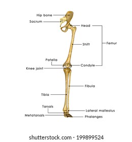
The femur is the single bone of the thigh. The foot bones shown in this diagram are the talus, navicular, cuneiform, cuboid, metatarsals and calcaneus. Please refer to the diagram for anatomical terms and splint summary. The bones of the foot are divided into three groups. The tibia and fibula are two long bones that run parallel to each other, forming the scaffold of the leg and providing attachment points for many muscles. There are in all 7 bones, which fall under tarsal bones category. Most leg pain results from wear and tear, overuse, or injuries in joints or bones or in muscles, ligaments, tendons or other soft tissues. Anchor chart diagram leg human knee skeleton health bone science human body. 6 10 2 votes muscle of the human leg diagram. Blood vessels and nerves enter the bone. The second largest bone in body is the tibia, also called the shinbone. The three bones which form the pelvis are the pubis, the ilium, and the ischium. The stifle joint connects the femur, which is the dog thigh bone, to the tibia and fibula, the lower leg bones, and the patella,the canine equivalent to the knee cap.
The major bones of the leg are the femur (thigh bone), tibia (shin bone), and adjacent fibula, and these are all long bones.the patella (kneecap) is the sesamoid bone in front of the knee.most of the leg skeleton has bony prominences and margins that can be palpated and some serve as anatomical landmarks that define the extent of the leg. Dog leg anatomy is complex, especially dog knees, which are found on the hind legs. The human leg consists of 8 bones, 4 per leg. Long bones are found in the arms (humerus, ulna, radius) and legs (femur, tibia, fibula), as well as in. The tarsal bones in the foot are located amongst tibia, metatarsal bones, and fibula.
Hip Thigh Atlas Of Anatomy from doctorlib.info Leg bones diagram femur manual e books. 6 10 2 votes muscle of the human leg diagram. Leg bones diagram diagram schematic ideas lower leg muscle diagram blank sketch coloring page antique 1890s medical anatomy diagram leg bones skeleton posted on april 18, 2019april 18, 2019. The ilium is the bone at the top of the waist, while the pubis bones are found just below the ilium. Editor · aug 13, 2017 ·. These landmarks are the anterior superior iliac spine. The foot bones shown in this diagram are the talus, navicular, cuneiform, cuboid, metatarsals and calcaneus. He leg's main function in the human is for locomotion and support of the rest of the body.
Leg pain can also be caused by blood clots, varicose veins or poor circulation.
Anatomically, the term leg means the part of the hind limb that extends from the stiffle joint to the hock joint (knee to ankle or tibia and fibula bones region). The knee joint is the largest joint in the body and is primarily a hinge joint, although some sliding and rotation occur. Related posts of diagram of leg bones inside of arm muscle and bone. Some types of leg pain can be traced to problems in your lower spine. The knee is a strong but flexible hinge joint that uses muscles and ligaments to withstand the torques and strains of powerful leg movements. 6 10 2 votes muscle of the human leg diagram. These landmarks are the anterior superior iliac spine. Posted on april 18, 2019april 18, 2019. The foot bones shown in this diagram are the talus, navicular, cuneiform, cuboid, metatarsals and calcaneus. The tibia and fibula are two long bones that run parallel to each other, forming the scaffold of the leg and providing attachment points for many muscles. Depending on the origin of the discomfort, upper leg pain symptoms can be a chronic nuisance or acute and debilitating. The upper leg, in particular, is comprised of bones and muscles that are susceptible to injury, particularly when excess strain is placed upon them. The diagram of bones in the ankle and foot is given below:
Ankle & lower leg anatomy. Long bones are found in the arms (humerus, ulna, radius) and legs (femur, tibia, fibula), as well as in. The tarsal bones in the foot are located amongst tibia, metatarsal bones, and fibula. The foot bones shown in this diagram are the talus, navicular, cuneiform, cuboid, metatarsals and calcaneus. These bones are arranged into two major divisions:
Labelled Bones Leg High Res Stock Images Shutterstock from image.shutterstock.com Next to the tibia is the fibula, the thinner,. The second largest bone in body is the tibia, also called the shinbone. Nerves leg diagram diagram continue reading. These bones are arranged into two major divisions: Some types of leg pain can be traced to problems in your lower spine. The technical term for a dog knee is the stifle joint. This long bone connects with the knee at one end and the ankle at the other. The foot bones shown in this diagram are the talus, navicular, cuneiform, cuboid, metatarsals and calcaneus.
Image result for leg bones diagram human leg bone structure your leg bones are the longest and strongest bones in your body.
Most leg pain results from wear and tear, overuse, or injuries in joints or bones or in muscles, ligaments, tendons or other soft tissues. The tarsal bones in the foot are located amongst tibia, metatarsal bones, and fibula. The bones of the foot are divided into three groups. With different grades of sprains depending on severity. Diagram of blood and nerve supply to bone. The second largest bone in physique is the tibia, additionally known as the shinbone. Bones and muscles anatomy 12 photos of the bones and muscles anatomy bones and muscles games, bones and muscles online games, bones and muscles ppt, bones and muscles used in a tennis serve, bones and muscles used in volleyball, bone, bones and muscles games, bones and muscles online games, bones and muscles ppt, bones. The bones of the leg are the femur, tibia, fibula and patella. The femur is the single bone of the thigh. Leg pain can also be caused by blood clots, varicose veins or poor circulation. The tibia, commonly known as the 'shin bone', is the largest and most medial of the two. The ischium is located just behind the pubis bone. Nerves leg diagram diagram continue reading.